|

| 产地 | 进口、国产 |
| 品牌 | 上海莼试 |
| 保存条件 | Store at -20 °C |
| 货号 | CS11903 |
| 应用范围 | WB=1:100-500 ELISA=1:500-1000 IP=1:20-100 IHC-P=1:100-500 IHC-F=1:100-500 IF=1:100-500 |
| CAS编号 | |
| 抗体名 | Anti-Phospho-Tie2 (Ser1119) |
| 克隆性 | |
| 靶点 | 详见说明书 |
| 适应物种 | 详见说明书 |
| 形态 | 详见说明书 |
| 宿主 | 详见说明书 |
| 亚型 | IgG |
| 标识物 | 详见说明书 |
| 浓度 | 1mg/1ml% |
| 免疫原 | KLH conjugated Synthesised phosphopeptide derived from human Tie2 around the phosphorylation site of Ser1119 |
产品订购信息:
英文名称 Anti-Phospho-Tie2 (Ser1119)
中文名称 磷酸化生成素受体2抗体品牌
别 名 Tie-2; Tie2; Tek; Angiopoietin-1 receptor; Tyrosine-protein kinase receptor TIE-2; hTIE2; Tyrosine-protein kinase receptor TEK; Tunica interna endothelial cell kinase; p140 TEK; Angiopoietin 1 receptor; CD202b; CD202b antigen; Endothelial tyrosine kinase; Endothelium specific receptor tyrosine kinase 2; hTIE 2; Hyk; Soluble TIE2 variant 1; Soluble TIE2 variant 2; tek tyrosine kinase; TEK tyrosine kinase endothelial; tek tyrosine kinase, endothelial; TIE 2; TIE2_HUMAN; Tunica interna endothelial cell kinase; Tyrosine kinase with Ig and EGF homology domains 2; Tyrosine protein kinase receptor TEK; Tyrosine protein kinase receptor TIE 2; Tyrosine-protein kinase receptor TIE-2; Venous malformations multiple cutaneous and mucosal; VMCM 1; VMCM; VMCM1; CD202b.


浓 度 1mg/1ml
规 格 0.1ml/100μg
抗体来源 Rabbit
克隆类型 polyclonal
交叉反应 Human, Mouse, Rat, Chicken, Dog, Pig, Cow, Horse, Rabbit, Sheep
产品类型 一抗 磷酸化抗体
研究领域 心 信号转导 干细胞 生长因子和激素 激酶和磷酸酶
蛋白分子量 predicted molecular weight: 124kDa
性 状 Lyophilized or Liquid
免 疫 原 KLH conjugated Synthesised phosphopeptide derived from human Tie2 around the phosphorylation site of Ser1119
亚 型 IgG
纯化方法 affinity purified by Protein A
储 存 液 0.01M PBS, pH 7.4 with 10 mg/ml BSA and 0.1% Sodium azide
磷酸化生成素受体2抗体品牌 产品应用 WB=1:100-500 ELISA=1:500-1000 IP=1:20-100 IHC-P=1:100-500 IHC-F=1:100-500 IF=1:100-500
(石蜡切片需做抗原修复)
not yet tested in other applications.
optimal dilutions/concentrations should be determined by the end user.
保存条件 Store at -20 °C for one year. Avoid repeated freeze/thaw cycles. The lyophilized antibody is stable at room temperature for at least one month and for greater than a year when kept at -20°C. When reconstituted in sterile pH 7.4 0.01M PBS or diluent of antibody the antibody is stable for at least two weeks at 2-4 °C.
Important Note This product as supplied is intended for research use only, not for use in human, therapeutic or diagnostic applications.
产品介绍 The TEK receptor tyrosine kinase is expressed almost exclusively in endothelial cells in mice, rats, and humans. This receptor possesses a unique extracellular domain containing 2 immunoglobulin-like loops separated by 3 epidermal growth factor-like repeats that are connected to 3 fibronectin type III-like repeats. The ligand for the receptor is angiopoietin-1. Defects in TEK are associated with inherited venous malformations; the TEK signaling pathway appears to be critical for endothelial cell-smooth muscle cell communication in venous morphogenesis.TEK is closely related to the TIE receptor tyrosine kinase.
Function : Tyrosine-protein kinase that acts as cell-surface receptor for ANGPT1, ANGPT2 and ANGPT4 and regulates angiogenesis, endothelial cell survival, proliferation, migration, adhesion and cell spreading, reorganization of the actin cytoskeleton, but also maintenance of vascular quiescence. Has anti-inflammatory effects by preventing the leakage of proinflammatory plasma proteins and leukocytes from blood vessels. Required for normal angiogenesis and heart development during embryogenesis. Required for post-natal hematopoiesis. After birth, activates or inhibits angiogenesis, depending on the context. Inhibits angiogenesis and promotes vascular stability in quiescent vessels, where endothelial cells have tight contacts. In quiescent vessels, ANGPT1 oligomers recruit TEK to cell-cell contacts, forming complexes with TEK molecules from adjoining cells, and this leads to preferential activation of phosphatidylinositol 3-kinase and the AKT1 signaling cascades. In migrating endothelial cells that lack cell-cell adhesions, ANGT1 recruits TEK to contacts with the extracellular matrix, leading to the formation of focal adhesion complexes, activation of PTK2/FAK and of the downstream kinases MAPK1/ERK2 and MAPK3/ERK1, and ultimately to the stimulation of sprouting angiogenesis. ANGPT1 signaling triggers receptor dimerization and autophosphorylation at specific tyrosine residues that then serve as binding sites for scaffold proteins and effectors. Signaling is modulated by ANGPT2 that has lower affinity for TEK, can promote TEK autophosphorylation in the absence of ANGPT1, but inhibits ANGPT1-mediated signaling by competing for the same binding site. Signaling is also modulated by formation of heterodimers with TIE1, and by proteolytic processing that gives rise to a soluble TEK extracellular domain. The soluble extracellular domain modulates signaling by functioning as decoy receptor for angiopoietins. TEK phosphorylates DOK2, GRB7, GRB14, PIK3R1; SHC1 and TIE1.
Subunit : Tyrosine-protein kinase that acts as cell-surface receptor for ANGPT1, ANGPT2 and ANGPT4 and regulates angiogenesis, endothelial cell survival, proliferation, migration, adhesion and cell spreading, reorganization of the actin cytoskeleton, but also maintenance of vascular quiescence. Has anti-inflammatory effects by preventing the leakage of proinflammatory plasma proteins and leukocytes from blood vessels. Required for normal angiogenesis and heart development during embryogenesis. Required for post-natal hematopoiesis. After birth, activates or inhibits angiogenesis, depending on the context. Inhibits angiogenesis and promotes vascular stability in quiescent vessels, where endothelial cells have tight contacts. In quiescent vessels, ANGPT1 oligomers recruit TEK to cell-cell contacts, forming complexes with TEK molecules from adjoining cells, and this leads to preferential activation of phosphatidylinositol 3-kinase and the AKT1 signaling cascades. In migrating endothelial cells that lack cell-cell adhesions, ANGT1 recruits TEK to contacts with the extracellular matrix, leading to the formation of focal adhesion complexes, activation of PTK2/FAK and of the downstream kinases MAPK1/ERK2 and MAPK3/ERK1, and ultimately to the stimulation of sprouting angiogenesis. ANGPT1 signaling triggers receptor dimerization and autophosphorylation at specific tyrosine residues that then serve as binding sites for scaffold proteins and effectors. Signaling is modulated by ANGPT2 that has lower affinity for TEK, can promote TEK autophosphorylation in the absence of ANGPT1, but inhibits ANGPT1-mediated signaling by competing for the same binding site. Signaling is also modulated by formation of heterodimers with TIE1, and by proteolytic processing that gives rise to a soluble TEK extracellular domain. The soluble extracellular domain modulates signaling by functioning as decoy receptor for angiopoietins. TEK phosphorylates DOK2, GRB7, GRB14, PIK3R1; SHC1 and TIE1.
Subcellular Location : Cell membrane; Single-pass type I membrane protein. Cell junction. Cell junction, focal adhesion. Cytoplasm, cytoskeleton. Secreted.
Tissue Specificity : Detected in umbilical vein endothelial cells. Proteolytic processing gives rise to a soluble extracellular domain that is detected in blood plasma (at protein level). Predominantly expressed in endothelial cells and their progenitors, the angioblasts. Has been directly found in placenta and lung, with a lower level in umbilical vein endothelial cells, brain and kidney.
Post-translational modifications : Proteolytic processing leads to the shedding of the extracellular domain (soluble TIE-2 alias sTIE-2).
Autophosphorylated on tyrosine residues in response to ligand binding. Autophosphorylation occurs in trans, i.e. one subunit of the dimeric receptor phosphorylates tyrosine residues on the other subunit. Autophosphorylation occurs in a sequential manner, where Tyr-992 in the kinase activation loop is phosphorylated first, followed by autophosphorylation at Tyr-1108 and at additional tyrosine residues. ANGPT1-induced phosphorylation is impaired during hypoxia, due to increased expression of ANGPT2. Phosphorylation is important for interaction with GRB14, PIK3R1 and PTPN11. Phosphorylation at Tyr-1102 is important for interaction with SHC1, GRB2 and GRB7. Phosphorylation at Tyr-1108 is important for interaction with DOK2 and for coupling to downstream signal transduction pathways in endothelial cells. Dephosphorylated by PTPRB.
Ubiquitinated. The phosphorylated receptor is ubiquitinated and internalized, leading to its degradation.
DISEASE : Defects in TEK are a cause of dominantly inherited venous malformations (VMCM) [MIM:600195]; an error of vascular morphogenesis characterized by dilated, serpiginous channels.
Note=May play a role in a range of diseases with a vascular component, including neovascularization of tumors, psoriasis and inflammation.
Similarity : Belongs to the protein kinase superfamily. Tyr protein kinase family. Tie subfamily.
Contains 3 EGF-like domains.
Contains 3 fibronectin type-III domains.
Contains 2 Ig-like C2-type (immunoglobulin-like)domains.
Contains 1 protein kinase domain.
Database links : UniProtKB/Swiss-Prot: Q02763.2
Tie2 是内皮特异性的酪氨酸激酶型受体, 主要表达在肺内皮以及卵泡、创口肉芽组织等内皮. 在发育中起重要的.

EPO human 人促红细胞生成素Multi-class antibodies
Anti-c-Myc tag c-Myc tag标签抗体Multi-class antibodies规格: 0.1ml
Rhesus antibody Rh NPAP1 核孔蛋白1抗体 规格 0.2ml
CD3/FITC + CD56/PE FITC标记CD3抗体 + PE标记CD56抗体 0.1mlx2
TSKS 英文名称: 特异激酶底物蛋白抗体 0.2ml
C6orf211 英文名称: 6号染色体开放阅读框211抗体 0.2ml
Anti-c-Myc tag c-Myc tag标签抗体Multi-class antibodies规格: 0.1ml
NGX6 (nasopharyngeal carcinoma/NPC associated gene 6) 细胞相关基因6(多肽)Multi-class antibodies规格: 0.5mg
Anti-APOJ 载脂蛋白J抗体Multi-class antibodies规格: 0.2ml
Rhesus antibody Rh Phospho-ATRIP (Ser224) 先关蛋白TREX1抗体 规格 0.1ml
兔抗山羊 IgG(H+L)/AP 0.1ml 美国Jackson公司分装
VEGF 英文名称: 内皮生长因子抗体 0.1ml
Desmoplakin I+II 英文名称: 桥粒斑蛋白1+2抗体 0.1ml
Anti-APOJ 载脂蛋白J抗体Multi-class antibodies规格: 0.2ml
Anti-SSEA4/FITC 荧光素标记阶段特异性胚胎表面抗原-4IgGMulti-class antibodies规格: 0.2ml
AMACR/P504S(alpha-methylacyl-CoA racemase) α-甲基酰基辅酶A消旋酶抗原Multi-class antibodies规格: 0.5mg
血小板源性生长因子受体-B抗体 Anti-PDGF-R-B 0.1ml
Secretogranin V 英文名称: 神经颗粒蛋白5抗体 0.2ml
ERAS 英文名称: 胚胎干细胞RAS蛋白抗体 0.2ml
Rhesus antibody Rh phospho-NDEL1(Ser242) 磷酸化中心粒蛋白Nudel抗体 规格 0.1ml
AMACR/P504S(alpha-methylacyl-CoA racemase) α-甲基酰基辅酶A消旋酶抗原Multi-class antibodies规格: 0.5mg
大鼠免疫球蛋白G(IgG)ELISA 试剂盒 96T/48T 试剂盒 组装/原装
人生长调节致癌基因α/黑素瘤生长刺激因子(GROα/CXCL1/MGSA)免疫试剂盒 Human GROα/MGSA ELISA Kit
Mouseglucocoicoidreceptor,GRELISAKit小鼠糖皮质类固醇受体(GR)ELISA试剂盒规格:96T/48T
Sophorajaponicaagglinin,SJAELISA试剂盒槐凝集素(SJA)ELISA试剂盒规格:96T/48T
HumanMaixmetalloproteinase10,MMP-10ELISA试剂盒人基质金属蛋白酶10(MMP-10)ELISA试剂盒规格:96T/48T
Humanphospholamban,PLNELISAKit人受磷蛋白(PLN)ELISA试剂盒规格:96T/48T
大鼠不对称二甲基精酸(ADMA)ELISA试剂盒 96T/48T 试剂盒 组装/原装
人免疫球蛋白E Fc段受体Ⅰ(FcεRⅠ)免疫试剂盒 Human Receptor Ⅰ for the Fc region of immunoglobulin E,FcεRⅠ ELISA Kit
MouseVascuoarendothelialcellgrowthfactorreceptor1,VEGFR-1/Flt1ELISAKit小鼠内皮细胞生长因子受体1(VEGFR-1/Flt1)ELISA试剂盒规格:96T/48T
石蜡切片组织CASPASE-3蛋白表达NBT显色光学显微镜检测试剂盒10/20次
humanProstaglandinF,PG-FELISA试剂盒人素F(PGF)ELISA试剂盒规格:96T/48T
HumahymosinELISAKit人胸腺肽(Thymosin)ELISA试剂盒规格:96T/48T
磷酸化生成素受体2抗体品牌 大鼠低氧诱导因子1α(HIF1α)ELISA试剂盒 ,英文名: HIF1α ELISA Kit
E selectin (E-Selectin/CD62E) ELISA Kit 人E选择素(E-Selectin/CD62E)ELISA试剂盒
Ratmonocytechemotacticprotein3,MCP-3ELISAkit 大鼠单核细胞趋化蛋白3(MCP-3/CCL7)ELISA试剂盒 96T/48T 进口分装
CLIAKitforCyPB(HumancyclophilinB)ELISAKit人嗜环蛋白/亲环素B规格:48T/96T
细胞色素P450亚酶CYP2A(COD)活性荧光定量检测试剂盒20次
Ratfreechorionicgonadoopin,f-βCGELISAKit大鼠游离β绒毛膜激素(f-βCG)ELISA试剂盒规格:96T/48T

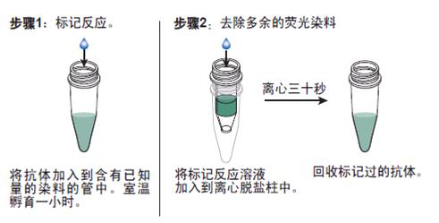
抗体的生物素化标记实验要点:
1. 磷酸化生成素受体2抗体品牌 如在反应混合液中有叠氮钠或游离氨基存在,会抑制标记反应。因此,蛋白质在反应前要对 0.1mol/L碳酸氢钠缓冲液或0.5mol/L硼酸缓冲液充分透析;
2.所用的NHSB及待生物素化蛋白质之间的分子比按蛋白质表面的ε-氨基的密度会有所不同,选择不当则影响标记的效率,应先用几个不同的分子比来筛选最适条件;
3.用NHSB量过量也是不利的,抗原的结合位点可能因此被封闭,导致抗体失活;
4.由于抗体的氨基不易接近可能造成生物素化不足,此时可加入去污剂如 Triton x-100, Tween20等;
5.当游离ε-氨基(赖氨酸残基的氨基)存在于抗体的抗原结合位点时,或位于酶的催化位点时,生物素化会降低或损伤抗体蛋白的结合力或活性;
6.生物素还可能与不同的功能基团,如羰基、氨基、巯基、异咪唑基及*基,也可与糖基共价结合;
7.交联反应后,应充分透析,否则,残余的生物素会对生物素化抗体与亲和素的结合产生竞争作用;
8.在细胞的荧光标记实验中,中和亲和素的本底低,但由于链霉亲和素含有少量正电荷,故对某些细胞可导致高本底。
抗体的鉴定:
1)磷酸化生成素受体2抗体品牌 抗体的效价鉴定:不管是用于诊断还是用于,制备抗体的目的都是要求较高效价。不同的抗原制备的抗体,要求的效价不一。鉴定效价的方法很多,包括有试管凝集反应,琼脂扩散试验,酶联免疫吸附试验等。常用的抗原所制备的抗体一般都有约成的鉴定效价的方法,以资比较。如制备抗抗体的效价,一般就采用琼脂扩散试验来鉴定。
2)抗体的特异性鉴定:抗体的特异性是指与相应抗原或近似抗原物质的识别能力。抗体的特异性高,它的识别能力就强。衡量特异性通常以交叉反应率来表示。交叉反应率可用竞争抑制试验测定。以不同浓度抗原和近似抗原分别做竞争抑制曲线,计算各自的结合率,求出各自在IC50时的浓度,并按公式计算交叉反应率。
如果所用抗原浓度IC50浓度为pg/管,而一些近似抗原物质的IC50浓度几乎是无穷大时,表示这一抗血清与其他抗原物质的交叉反应率近似为0,即该血清的特异性较好。
3)抗体亲和力:是指抗体和抗原结合的牢固程度。亲和力的高低是由抗原分子的大小,抗体分子的结合位点与抗原决定簇之间立体构型的合适度决定的。有助于维持抗原抗体复合物稳定的分子间力有氢键,疏水键,侧链相反电荷基因的库仑力,范德华力和空间斥力。亲和力常以亲和常数K表示,K的单位是L/mol。抗体亲和力的测定对抗体的筛选,确定抗体的用途,验证抗体的均一性等均有重要意义。