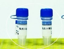
|

| 产地 | 进口、国产 |
| 品牌 | 上海莼试 |
| 保存条件 | Store at -20 °C |
| 货号 | CS11180 |
| 应用范围 | WB=1:100-500 ELISA=1:500-1000 IP=1:20-100 IHC-P=1:100-500 IHC-F=1:100-500 IF=1:100-500 |
| CAS编号 | |
| 抗体名 | Anti-Phospho-Rb/P105 RB (Thr821) |
| 克隆性 | |
| 靶点 | 详见说明书 |
| 适应物种 | 详见说明书 |
| 形态 | 详见说明书 |
| 宿主 | 详见说明书 |
| 亚型 | IgG |
| 标识物 | 详见说明书 |
| 浓度 | 1mg/1ml% |
| 免疫原 | KLH conjugated Synthesised phosphopeptide derived from human Retinoblastoma associated protein around the phosphorylation site of Thr821 |
产品订购信息:
英文名称 Anti-Phospho-Rb/P105 RB (Thr821)
中文名称 磷酸化视网膜母细胞瘤相关蛋白1抗体说明书
别 名 OSRC; P105 RB; P105RB; PP105; PP110; pRb; RB 1; RB1; RB1 protein; Retinoblastoma 1 (including osteosarcoma); Retinoblastoma 1; Retinoblastoma associated protein; Retinoblastoma related osteosarcoma; Retinoblastoma susceptibility gene; Including osteosarcoma; RB_HUMAN.


浓 度 1mg/1ml
规 格 0.1ml/100μg
抗体来源 Rabbit
克隆类型 polyclonal
交叉反应 Human, Mouse, Rat, Pig, Cow, Horse, Rabbit
产品类型 一抗 磷酸化抗体
研究领域 细胞生物 免疫学 信号转导 转录调节因子
蛋白分子量 predicted molecular weight: 102kDa
性 状 Lyophilized or Liquid
免 疫 原 KLH conjugated Synthesised phosphopeptide derived from human Retinoblastoma associated protein around the phosphorylation site of Thr821
亚 型 IgG
纯化方法 affinity purified by Protein A
储 存 液 0.01M PBS, pH 7.4 with 10 mg/ml BSA and 0.1% Sodium azide
磷酸化视网膜母细胞瘤相关蛋白1抗体说明书 产品应用 WB=1:100-500 ELISA=1:500-1000 IP=1:20-100 IHC-P=1:100-500 IHC-F=1:100-500 IF=1:100-500
(石蜡切片需做抗原修复)
not yet tested in other applications.
optimal dilutions/concentrations should be determined by the end user.
保存条件 Store at -20 °C for one year. Avoid repeated freeze/thaw cycles. The lyophilized antibody is stable at room temperature for at least one month and for greater than a year when kept at -20°C. When reconstituted in sterile pH 7.4 0.01M PBS or diluent of antibody the antibody is stable for at least two weeks at 2-4 °C.
Important Note This product as supplied is intended for research use only, not for use in human, therapeutic or diagnostic applications.
产品介绍 Rb is a tumor suppressor gene which functions as a negative regulator of the cell cycle by interacting with transcription factors including E2F1, PU1, ATF2, UBF, Elf1 and cAbl. This ability of Rb to alter transcription is regulated by phosphorylation catalyzed by the cyclin dependent protein kinases (cdks). Rb is phosphorylated on serine and threonine, but not on tyrosine residues. It forms a complex with SV40 large T antigen, adenovirus E1A, and human papilloma virus 16E. Rb protein may act by regulating transcription and loss of its function leads to uncontrolled cell growth. Aberrations in the Rb gene have been implicated in cancers of breast, colon, prostate, kidney, nasopharynx, and leukemia.
Function : Key regulator of entry into cell division that acts as a tumor suppressor. Promotes G0-G1 transition when phosphorylated by CDK3/cyclin-C. Acts as a transcription repressor of E2F1 target genes. The underphosphorylated, active form of RB1 interacts with E2F1 and represses its transcription activity, leading to cell cycle arrest. Directly involved in heterochromatin formation by maintaining overall chromatin structure and, in particular, that of constitutive heterochromatin by stabilizing histone methylation. Recruits and targets histone methyltransferases SUV39H1, SUV420H1 and SUV420H2, leading to epigenetic transcriptional repression. Controls histone H4 'Lys-20' trimethylation. Inhibits the intrinsic kinase activity of TAF1. Mediates transcriptional repression by SMARCA4/BRG1 by recruiting a histone deacetylase (HDAC) complex to the c-FOS promoter. In resting neurons, transcription of the c-FOS promoter is inhibited by BRG1-dependent recruitment of a phospho-RB1-HDAC1 repressor complex. Upon calcium influx, RB1 is dephosphorylated by calcineurin, which leads to release of the repressor complex (By similarity). In case of viral infections, interactions with SV40 large T antigen, HPV E7 protein or adenovirus E1A protein induce the disassembly of RB1-E2F1 complex thereby disrupting RB1's activity.
Subunit : Interacts with ATAD5. Interacts with PRMT2, CDK1 and CDK2. The hypophosphorylated form interacts with and sequesters the E2F1 transcription factor. Interacts with heterodimeric E2F/DP transcription factor complexes containing TFDP1 and either E2F1, E2F3, E2F4 or E2F5, or TFDP2 and E2F4. The unphosphorylated form interacts with EID1, ARID3B, KDM5A, SUV39H1, MJD2A/JHDM3A and THOC1. Interacts with the N-terminal domain of TAF1. Interacts with AATF, DNMT1, LIN9, LMNA, SUV420H1, SUV420H2, PELP1 and TMPO-alpha. May interact with NDC80. Interacts with GRIP1 and UBR4. Interacts with ARID4A and KDM5B. Interacts with E4F1 and LIMD1. Interacts with SMARCA4/BRG1 AND HDAC1. Interacts with adenovirus E1A protein, HPV E7 protein and SV40 large T antigen. Interacts with PSMA3 and USP4. Interacts (when methylated at Lys-860) with L3MBTL1.
Subcellular Location : Nucleus.
Tissue Specificity : Expressed in the retina.
Post-translational modifications : Phosphorylated by CDK6 and CDK4, and subsequently by CDK2 at Ser-567 in G1, thereby releasing E2F1 which is then able to activate cell growth. Dephosphorylated at the late M phase. SV40 large T antigen, HPV E7 and adenovirus E1A bind to the underphosphorylated, active form of pRb. Phosphorylation at Thr-821 and Thr-826 promotes interaction between the C-terminal domain C and the Pocket domain, and thereby inhibits interactions with heterodimeric E2F/DP transcription factor complexes. Dephosphorylated at Ser-795 by calcineruin upon calcium stimulation. CDK3/cyclin-C-mediated phosphorylation at Ser-807 and Ser-811 is required for G0-G1 transition. Phosphorylated by CDK1 and CDK2 upon TGFB1-mediated apoptosis.
N-terminus is methylated by METTL11A/NTM1. Monomethylated at Lys-860 by SMYD2, promoting interaction with L3MBTL1.
DISEASE : Defects in RB1 are the cause of childhood cancer retinoblastoma (RB) [MIM:180200]. RB is a congenital malignant tumor that arises from the nuclear layers of the retina. It occurs in about 1:20'000 live births and represents about 2% of childhood malignancies. It is bilateral in about 30% of cases. Although most RB appear sporadically, about 20% are transmitted as an autosomal dominant trait with incomplete penetrance. The diagnosis is usually made before the age of 2 years when strabismus or a gray to yellow reflex from pupil ('cat eye') is investigated.
Defects in RB1 are a cause of susceptibility to bladder cancer (BLC) [MIM:109800]. A malignancy originating in tissues of the urinary bladder. It often presents with multiple tumors appearing at different times and at different sites in the bladder. Most bladder cancers are transitional cell carcinomas. They begin in cells that normally make up the inner lining of the bladder. Other types of bladder cancer include squamous cell carcinoma (cancer that begins in thin, flat cells) and adenocarcinoma (cancer that begins in cells that make and release mucus and other fluids). Bladder cancer is a complex disorder with both genetic and environmental influences.
Defects in RB1 are a cause of osteogenic sarcoma (OSRC) [MIM:259500].
Similarity : Belongs to the retinoblastoma protein (RB) family.
Database links : UniProtKB/Swiss-Prot: P06400.2

MIP2 (myocardial ischemic preconditioning upregulated protein 2) 巨噬细胞蛋白2抗原Multi-class antibodies规格: 0.5mg
Anti-Angiopoietin 1 生成素-1抗体Multi-class antibodies规格: 0.1ml
Rhesus antibody Rh phospho-Beta-catenin(Tyr86) 磷酸化β 连环素蛋白抗体 规格 0.1ml
包被缓冲液(PH9.6) 100ml 进口分装
VTG 英文名称: 鱼、青鳉鱼卵黄蛋白原抗体 0.2ml
DLGAP4 英文名称: PSD95结合蛋白4抗体 0.2ml
Anti-Angiopoietin 1 生成素-1抗体Multi-class antibodies规格: 0.1ml
Anti-phospho-P53(pSer392)/FITC 荧光素标记抗磷酸化抑制基因P53抗体IgGMulti-class antibodies规格: 0.2ml
Rabbit Anti-mouse IgG/Gold 金标记兔抗小鼠IgG(10nm/15nm)Multi-class antibodies规格: 0.5ml
纤维连结蛋白抗体 Anti-FN 0.1ml
Goat Anti-Mouse IgG/Alexa Fluor 350 Alexa Fluor 350标记的羊抗小鼠IgG 0.1ml
FNDC4 英文名称: Ⅲ型纤维连接蛋白域蛋白4抗体 0.2ml
Rhesus antibody Rh phospho-STAT5a(Tyr694) 磷酸化信号转导和转录激活因子5a抗体 规格 0.1ml
Rabbit Anti-mouse IgG/Gold 金标记兔抗小鼠IgG(10nm/15nm)Multi-class antibodies规格: 0.5ml
Syntenin 1 英文名称: 多配体聚糖结合蛋白1抗体 0.2ml
Endostatin 英文名称: 内皮抑素/内皮他丁抗体 0.1ml
组织蛋白酶G抗体 Anti-CTSG/CG 0.1ml
Anti-UCP-3/FITC 荧光素标记线粒体脱偶连蛋白3抗体IgGMulti-class antibodies规格: 0.2ml
Rhesus antibody Rh Phospho-Histone H3(Ser28) 磷酸化组蛋白H3抗体 规格 0.1ml
CXCR6(CXC-chemokine receptor 6) peptide 细胞表面趋化因子受体6抗原Multi-class antibodies规格: 0.5mg
RDFs, 大鼠真皮成纤维细胞
16HBE 人上皮细胞
MIF Others Mouse 小鼠 MIF / Migration Inhibitory Factor 人细胞裂解液 (阳性对照)
CL-0263BGC-803(人细胞)5×10⁶cells/瓶×2
人肝内胆管上皮细胞cDNAHIBEpiC cDNA
Asp2人胚胎肾细胞转化细胞;FC33 人肾小球内皮细胞完全培养基 100mL
CL-0148MADB106(大鼠癌细胞)5×10⁶cells/瓶×2
SELL Others Cynomolgus 食蟹猴 CD62L / L-Selectin / SELL 人细胞裂解液 (阳性对照)
人上皮细胞裂解物HPEpiCL
1590细胞,人癌细胞系 小鼠,EL-4细胞 脑平滑肌细胞Many types of cells包装:5 × 10⁵次方(1ml)
Beta-TC-6(小鼠胰岛素瘤胰岛β细胞) 5×10⁶cells/瓶×2
HCASMC Pellet 人平滑肌细胞团块 > 1 mio.cells 人成纤维细胞-心室裂解物HCF-av L
磷酸化视网膜母细胞瘤相关蛋白1抗体说明书 RDFs, 大鼠真皮成纤维细胞
16HBE 人上皮细胞
MIF Others Mouse 小鼠 MIF / Migration Inhibitory Factor 人细胞裂解液 (阳性对照)
CL-0263BGC-803(人细胞)5×10⁶cells/瓶×2
人肝内胆管上皮细胞cDNAHIBEpiC cDNA
Asp2人胚胎肾细胞转化细胞;FC33 人肾小球内皮细胞完全培养基 100mL

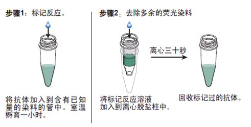
抗体的生物素化标记实验要点:
1. 磷酸化视网膜母细胞瘤相关蛋白1抗体说明书 如在反应混合液中有叠氮钠或游离氨基存在,会抑制标记反应。因此,蛋白质在反应前要对 0.1mol/L碳酸氢钠缓冲液或0.5mol/L硼酸缓冲液充分透析;
2.所用的NHSB及待生物素化蛋白质之间的分子比按蛋白质表面的ε-氨基的密度会有所不同,选择不当则影响标记的效率,应先用几个不同的分子比来筛选最适条件;
3.用NHSB量过量也是不利的,抗原的结合位点可能因此被封闭,导致抗体失活;
4.由于抗体的氨基不易接近可能造成生物素化不足,此时可加入去污剂如 Triton x-100, Tween20等;
5.当游离ε-氨基(赖氨酸残基的氨基)存在于抗体的抗原结合位点时,或位于酶的催化位点时,生物素化会降低或损伤抗体蛋白的结合力或活性;
6.生物素还可能与不同的功能基团,如羰基、氨基、巯基、异咪唑基及苯酚基,也可与糖基共价结合;
7.交联反应后,应充分透析,否则,残余的生物素会对生物素化抗体与亲和素的结合产生竞争作用;
8.在细胞的荧光标记实验中,中和亲和素的本底低,但由于链霉亲和素含有少量正电荷,故对某些细胞可导致高本底。
抗体的鉴定:
1)磷酸化视网膜母细胞瘤相关蛋白1抗体说明书 抗体的效价鉴定:不管是用于诊断还是用于,制备抗体的目的都是要求较高效价。不同的抗原制备的抗体,要求的效价不一。鉴定效价的方法很多,包括有试管凝集反应,琼脂扩散试验,酶联免疫吸附试验等。常用的抗原所制备的抗体一般都有约成的鉴定效价的方法,以资比较。如制备抗抗体的效价,一般就采用琼脂扩散试验来鉴定。
2)抗体的特异性鉴定:抗体的特异性是指与相应抗原或近似抗原物质的识别能力。抗体的特异性高,它的识别能力就强。衡量特异性通常以交叉反应率来表示。交叉反应率可用竞争抑制试验测定。以不同浓度抗原和近似抗原分别做竞争抑制曲线,计算各自的结合率,求出各自在IC50时的浓度,并按公式计算交叉反应率。
如果所用抗原浓度IC50浓度为pg/管,而一些近似抗原物质的IC50浓度几乎是无穷大时,表示这一抗血清与其他抗原物质的交叉反应率近似为0,即该血清的特异性较好。
3)抗体亲和力:是指抗体和抗原结合的牢固程度。亲和力的高低是由抗原分子的大小,抗体分子的结合位点与抗原决定簇之间立体构型的合适度决定的。有助于维持抗原抗体复合物稳定的分子间力有氢键,疏水键,侧链相反电荷基因的库仑力,范德华力和空间斥力。亲和力常以亲和常数K表示,K的单位是L/mol。抗体亲和力的测定对抗体的筛选,确定抗体的用途,验证抗体的均一性等均有重要意义。